Heights of Hyperkalemia
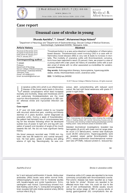
At 2 am, a 67 year old man, Hypertensive and Diabetic since 20 years, already a known case of Chronic Kidney Disease on Maintenance Hemodialysis since 10 years presented 1 month late for his Hemodialysis, came with the complains of Facial Puffiness and Abdominal distension since 1 month and also told that his bilateral pedal edema had progressed upto his thighs over the last 2 weeks. Dyspnea grade 4 since 2 days, Reduced urine output since 2 days.


He was Pale with pitting type of pedal edema.
His Respiratory rate was 27 cpm, Pulse rate of 100 bpm, Blood Pressure of 70/60mmhg with a Saturation of 79 % on Room Air.
On Auscultation of his lungs fields, there were Bilateral coarse inspiratory crackles all over his lung fields.
Heart sounds S1,S2 could be auscultated.
His Abdomen was distended.
Patient was put on Oxygen supplementation
NORADRENALINE infusion was started at 8 mcg/min

His ECG showed Sinus tachycardia with no P waves, Wide QRS complex of 200ms, Tall T waves - ?Due to Hyperkalemia
His ABG showed a pH of 7.1, PCO2 - 20, PO2 - 124, HCO3 - 5.5
showing severe metabolic acidosis with compensatory respiratory alkalosis.
Gave 10 ml of 10% calcium gluconate infusion IV over 10 minutes for hyperkalemia
10 units of insulin in 25% dextrose was started intravenously
and Salbutamol inhalation of 20mg in 4ml NS was given.
'
After 30 minutes, the ecg showed no significant changes, another infusion of 10ml of Calcium Gluconate was given over 10 minutes.
Calcium Gluconate reduces the membrane excitability reducing the potassium toxic effects on myocardium, it doesn't reduce the Serum levels of potassium as such. Whereas Insulin and Salbutamol shift potassium into the cells reducing serum potassium levels.

Chest Xray PA View suggestive of Pulmonary Edema - Batwing appearance
His Hemogram revealed a Hemoglobin of 7 g/dl, TLC - 17,000 cells/cumm, Platelets - 1.70 L/cumm
Serum Creatinine - 20mg/dl, Blood Urea - 467 mg/dl, Serum Potassium of 9 mEQ/L.
Diagnosis - Hyperkalemia with Metabolic Acidosis
? Septic Shock
Fluid overload secondary to Chronic Renal Failure
(CKD secondary to Hypertension & Diabetes Mellitus type 2 over 10 yrs, Chronic Alcoholic and Tobacco smoker since 25 years)
The patient was immediately taken up for hemodialysis.
ECG CHANGES IN HYPERKALEMIA

The earliest sign in ECG showing Hyperkalemia are the Tall peaked T waves when Serum Potassium is above 5.5 mEq/L
followed by loss of P waves and Widening of QRS complexes
When the Serum Potassium is >8mEq/L , sine waves are seen which occur due to fusion of QRS complexes with T waves in the absence of P waves.
> 9 mEq/L - Atrioventricular dissociation, ventricular tachycardias are seen.
Increase in potassium levels causes decrease in cardiac conduction leading to Increase in PR interval, Increase in QRS complex duration, reduced P wave amplitude eventually leading to absent P waves.
Absent P waves does not suggest that there is a sinus arrest, the SA node continues to work causing ventricular contraction rather than atrial contraction. This rhythm is known as Sinoventricular rhythm.
When potassium level increases further, the QRS complexes merge with T waves in the absence P wave showing a sine wave pattern.


Comments
Post a Comment